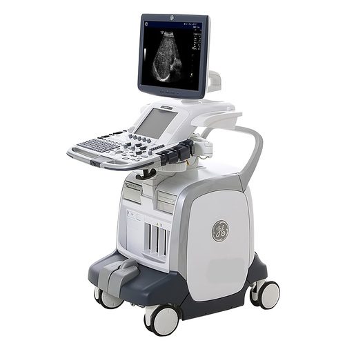
GE Logiq™ S8 Ultrasound system with B-Flow Imaging is available for rent and sale from US Med-Equip.
The B-Flow* is a blood flow visualization technique that displays the blood flow echoes in gray scale imaging, with different gray intensities according to the reflectors speed and dynamics. Based on the GE-patented Digitally Encoded Ultrasound technique to digitally suppress unwanted signals (e.g. noise and tissue) and boost weak signals (e.g. blood echoes), B-Flow overcomes the limitations of Doppler with the following imaging advantages:
• Direct hemodynamics visualization
• No vessel wall overlap (no overlay technique)
• Less dependency on the user or scanning angle
• Higher frame rate and spatial resolution than Color Flow
B-Flow Color
While B-Flow passes through the Color Processing channel (with exception of the Color Doppler Process), B-Flow Color can be displayed within a selected ROI with the following additional benefits:
• Easy display of small vessels
• Simultaneous B-Mode and B-Flow Color visualization
• Separate settings from B-Mode • Less tissue motion artifacts B-Flow and B-Flow Color features
• Dual or single Display
• ON/OFF tissue background information and B-Flow
• B-Flow or B-Flow Color selection
• Accumulation Mode, adding multiple frames
• Working with PW for flow quantification
• Easy 3D B-Flow imaging
B-Flow may help visualize
• Vessel-wall irregularities
• Stenosis with measurement
• Carotid plaque for vulnerability study (e.g. ulceration)
• Interaction of blood flow with anatomical structures inside the vessel such as venous valve cusps and thrombi
• Grafts for monitoring (e.g dialysis graft pseudoaneurysms)
• Thyroid nodule activity for assessment and monitoring
• Kidney perfusion (e.g. after transplants)
• Vascular disease after transfemoral catheterization (e.g. Aneurysm spurium, AV Fistula, Dissections, Hematomas, etc.)
• Liver and spleen vasculature
• Bladder reflux or jets
• Neonatal head vessels
• Cardiac Septal Defects (e.g. PFO, VSD, ASD, etc.)
• Endocardial walls in difficult to image patients